UNIT 4 HW & Quizzes Get a hint What are the structures included in the heart’s conduction system? Check all that apply. -Purkinje fibers -Atrioventricular bundle -atrioventricular (AV) node -Sinoatrial (SA) node Click the card to flip 👆 -Purkinje fibers -Atrioventricular bundle -atrioventricular (AV) node -Sinoatrial (SA) node
Structure and Function of the Heart
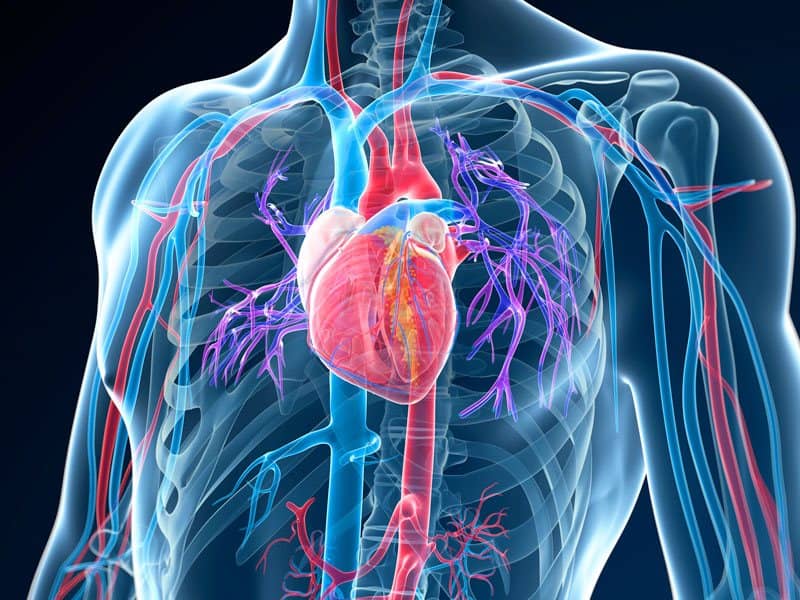
Which of the following structures is located in the middle mediastinum? (A) thoracic duct (B) lungs (C) esophagus(D) heart (E) azygos vein: 13. All of the following statements correctly apply to the right atrium EXCEPT (A) It receives blood from the superior and inferior vena cava and coronary sinus. (B) It forms the right side of the heart.

Source Image: chegg.com
Download Image
Sep 19, 2023Thoracic wall The first step in understanding thorax anatomy is to find out its boundaries. The thoracic, or chest wall, consists of a skeletal framework, fascia, muscles, and neurovasculature – all connected together to form a strong and protective yet flexible cage.. The thorax has two major openings: the superior thoracic aperture found superiorly and the inferior thoracic aperture

Source Image: dyhpoon.com
Download Image
FREE KS2 Simple Heart Diagram to label by PlanBee Chapter 20 Blood Vessels Terms in this set (75) Drag each image on the left to the type of vessel it represents on the right. Correctly label the following vessels and chemoreceptors in the superior portion of the heart. Correctly label the anatomical features of a continuous capillary.

Source Image: cardiacwellnessinstitute.com
Download Image
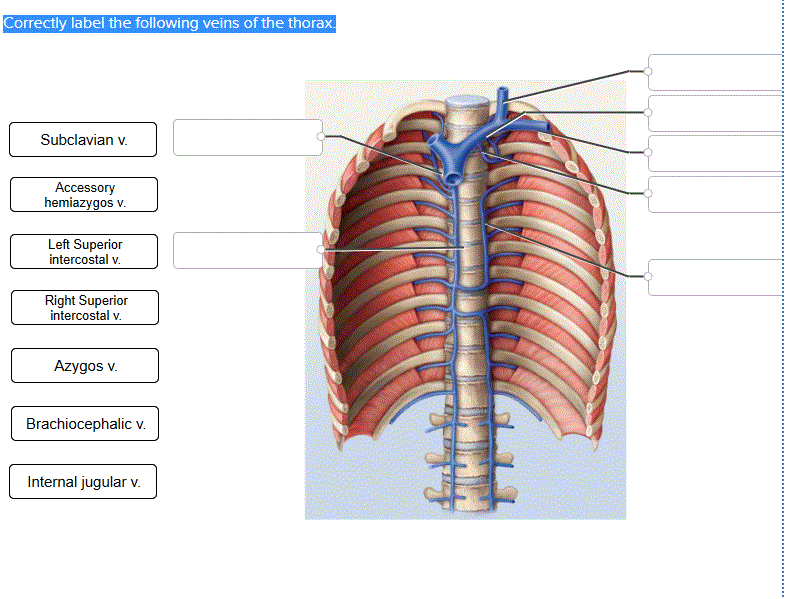
Correctly Label The Following Veins Of The Thorax.
Chapter 20 Blood Vessels Terms in this set (75) Drag each image on the left to the type of vessel it represents on the right. Correctly label the following vessels and chemoreceptors in the superior portion of the heart. Correctly label the anatomical features of a continuous capillary. 7. Systemic Veins: Bring blood back toward the heart. 8. Venae Cavae: The two large veins that return blood to the right atrium. Click the card to flip 👆 1 / 71 Flashcards Learn Test Match Q-Chat Created by Alexander_Pena20 Students also viewed A&P – Anatomy & Physiology: The Unity of Form and Function – Chapter 20: The Blood Vessels – MEGAKIT
Cardiac Wellness Institute – Official Blog
Jul 30, 2023Introduction. The thorax is the region between the abdomen inferiorly and the root of the neck superiorly. [1] [2] The thorax forms from the thoracic wall, its superficial structures (breast, muscles, and skin), and the thoracic cavity. A thorough comprehension of the anatomy and function of the thorax will help identify, differentiate, and Deep vein thrombosis (DVT) – symptoms, signs and treatment | healthdirect

Source Image: healthdirect.gov.au
Download Image
Can You Have a Pulmonary Embolism and Not Know It? Jul 30, 2023Introduction. The thorax is the region between the abdomen inferiorly and the root of the neck superiorly. [1] [2] The thorax forms from the thoracic wall, its superficial structures (breast, muscles, and skin), and the thoracic cavity. A thorough comprehension of the anatomy and function of the thorax will help identify, differentiate, and

Source Image: centerforvein.com
Download Image
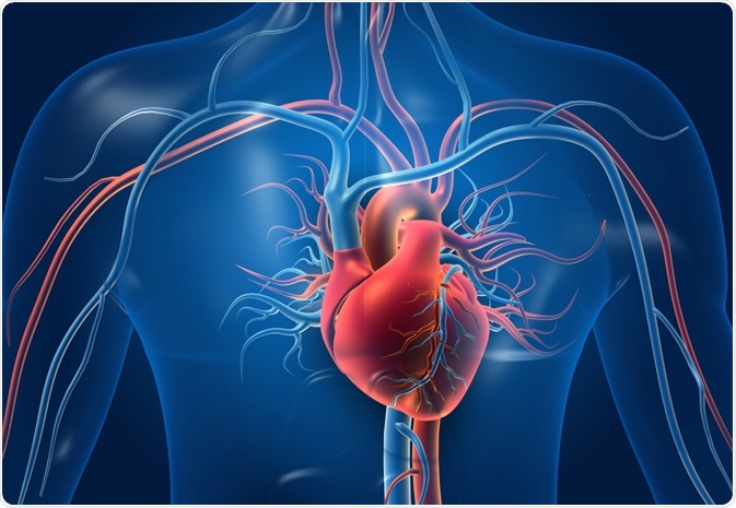
Structure and Function of the Heart UNIT 4 HW & Quizzes Get a hint What are the structures included in the heart’s conduction system? Check all that apply. -Purkinje fibers -Atrioventricular bundle -atrioventricular (AV) node -Sinoatrial (SA) node Click the card to flip 👆 -Purkinje fibers -Atrioventricular bundle -atrioventricular (AV) node -Sinoatrial (SA) node

Source Image: news-medical.net
Download Image
FREE KS2 Simple Heart Diagram to label by PlanBee Sep 19, 2023Thoracic wall The first step in understanding thorax anatomy is to find out its boundaries. The thoracic, or chest wall, consists of a skeletal framework, fascia, muscles, and neurovasculature – all connected together to form a strong and protective yet flexible cage.. The thorax has two major openings: the superior thoracic aperture found superiorly and the inferior thoracic aperture

Source Image: planbee.com
Download Image
Evaluating the Heart Size on Radiographs – Veterinary Medicine at Illinois Tributaries are the left vertebral, internal thoracic, inferior thyroid, superior intercostal, thymic vein, and pericardiophrenic veins. Variations of the brachiocephalic veins include entering the right atrium separately and the configuration of a left vena cava. Brachiocephalic Veins Internal Thoracic Veins (Mammary) (Fig. 8.8)

Source Image: vetmed.illinois.edu
Download Image
CT Scans and Blocked Arteries – PDC Chapter 20 Blood Vessels Terms in this set (75) Drag each image on the left to the type of vessel it represents on the right. Correctly label the following vessels and chemoreceptors in the superior portion of the heart. Correctly label the anatomical features of a continuous capillary.

Source Image: pdcenterlv.com
Download Image
Does central line position matter? Can we use ultrasonography to confirm line position? 7. Systemic Veins: Bring blood back toward the heart. 8. Venae Cavae: The two large veins that return blood to the right atrium. Click the card to flip 👆 1 / 71 Flashcards Learn Test Match Q-Chat Created by Alexander_Pena20 Students also viewed A&P – Anatomy & Physiology: The Unity of Form and Function – Chapter 20: The Blood Vessels – MEGAKIT

Source Image: emcrit.org
Download Image
Can You Have a Pulmonary Embolism and Not Know It?
Does central line position matter? Can we use ultrasonography to confirm line position? Which of the following structures is located in the middle mediastinum? (A) thoracic duct (B) lungs (C) esophagus(D) heart (E) azygos vein: 13. All of the following statements correctly apply to the right atrium EXCEPT (A) It receives blood from the superior and inferior vena cava and coronary sinus. (B) It forms the right side of the heart.
FREE KS2 Simple Heart Diagram to label by PlanBee CT Scans and Blocked Arteries – PDC Tributaries are the left vertebral, internal thoracic, inferior thyroid, superior intercostal, thymic vein, and pericardiophrenic veins. Variations of the brachiocephalic veins include entering the right atrium separately and the configuration of a left vena cava. Brachiocephalic Veins Internal Thoracic Veins (Mammary) (Fig. 8.8)